





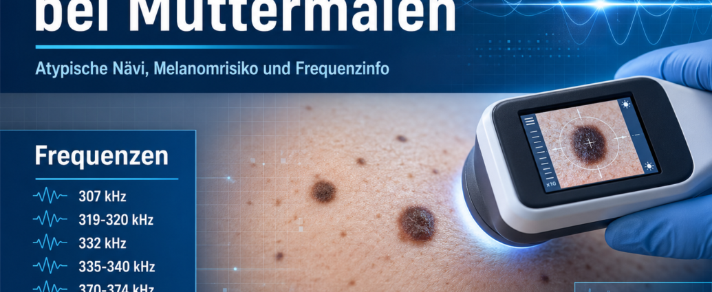
The Frequency therapy in the case of moles is described in the complementary context as a supplementary consideration to conventional medical principles, atypical nevi, genetic risk factors, UV exposure, melanoma risk, and possible resonance patterns. Moles, also known as nevi, are pigmented skin lesions that can arise from melanocytes or clusters of melanocytes. Most moles are benign, but certain atypical or dysplastic forms may require special attention.
The term „moles“ is used in the literature to describe various clinical conditions. On the one hand, it refers to trophoblastic changes following abnormal fertilization, such as complete and partial hydatidiform moles, invasive moles, choriocarcinomas, and trophoblastic tumors of the placental site. On the other hand, the literature describes atypical moles and dysplastic nevi of the skin. This article focuses on the dermatological significance of moles, atypical nevi, and their relationship to Melanoma in the foreground.
Frequency Therapy for Moles: An Overview from Conventional Medicine
Moles are common skin lesions. They can be present at birth or develop over the course of a person’s life. Many moles remain unchanged and harmless. Others may change in shape, color, size, or border and should be examined by a dermatologist.
Atypical moles differ from ordinary acquired nevi in certain clinical characteristics. These include a larger diameter, uneven pigmentation, irregular borders, or an irregular appearance. Some atypical moles may resemble melanoma and cannot always be reliably distinguished by their appearance alone.
Dysplastic nevi are moles that show histological abnormalities. The terms “atypical nevus,” “dysplastic nevus,” and “clinically suspicious mole” are used interchangeably in the literature. As a result, the distinction between them is not always consistent. Therefore, a precise dermatological evaluation—and, if necessary, a histological examination—is crucial.
Frequency Therapy for Moles and Atypical Nevi
Atypical nevi are acquired melanocytic skin lesions. They can occur singly or in large numbers. Clinically, they may be noticeable due to their irregular shape, varying shades of color, and larger size.
Not every conspicuous mole is dangerous. At the same time, some moles that appear unremarkable on the surface may show histological changes. The literature describes that clinical and histological assessments do not always fully correspond. A birthmark may therefore appear suspicious externally without exhibiting clear dysplastic features. Conversely, seemingly normal nevi may reveal histologically abnormal structures.
This uncertainty explains why suspicious skin lesions should not be assessed based solely on visual inspection. Dermoscopy, follow-up monitoring, and tissue examination are important components of the medical evaluation.
Atypical Moles and Melanoma Risk
Atypical moles are of particular interest because, in some cases, they can be difficult to distinguish clinically from malignant melanoma. Melanoma is a malignant Tumor the pigment-producing melanocytes and can metastasize early.
The literature describes how atypical moles are often thought to be associated with the melanoma virus. In the context of complementary frequency theory, therefore, certain resonances are examined that are cited in connection with both moles and melanomas.
People with many moles, conspicuous pigmented lesions, a family history of melanoma, or a previous diagnosis of melanoma deserve special attention. These individuals may be at increased risk for further conspicuous skin changes.
Frequency Therapy for Moles and Family History
Atypical moles can occur sporadically or run in families. The literature describes familial atypical nevus and melanoma syndrome, in which atypical nevi and melanomas occur with increased frequency in families.
The clinical and histological characteristics of familial atypical moles may overlap with those of sporadic atypical moles. Therefore, a family history is important. If several close relatives have had melanoma or if there is a notably high number of atypical nevi, a particularly thorough skin examination should be performed.
In complementary frequency therapy, a family history of the condition is not considered in isolation. It is evaluated in conjunction with skin type, UV exposure, immune status, pigment behavior, individual regulatory capacity, and possible resonance patterns.
Genetic Factors in Moles and Melanoma
The literature mentions various genes associated with hereditary forms of melanoma and atypical moles. These include CDKN2A, CDK4, and CMM1. These genes are linked to certain hereditary predispositions to melanoma and to familial atypical mole and melanoma syndrome.
Somatic mutations in genes such as PTEN, BRAF, and MC1R have also been described in connection with melanoma. MC1R, the melanocortin-1 receptor, is associated with pigmentation, skin type, and UV sensitivity.
These genetic factors do not automatically mean that melanoma will develop. However, they can influence an individual’s susceptibility to UV light, changes in pigmentation, and tumor development.
Frequency Therapy for Moles and UV Light
UV light is described in the literature as an important factor in the transformation of melanocytes into atypical melanocytes or melanoma cells. Both UVA and UVB radiation can influence biological processes in the skin.
UV radiation can cause DNA damage, promote inflammatory reactions, and disrupt cell regulation. Repeated sunburns, intense sun exposure, and inadequate sun protection, in particular, can take a toll on the skin over the long term.
For people with a family history of melanoma, UV light can be particularly significant. The literature indicates that UV exposure may be necessary for certain hereditary risk syndromes to fully manifest. For this reason, skin protection, regular checkups, and early evaluation of any suspicious changes are especially important.
Clinical Characteristics of Suspicious Moles
Unusual moles can look very different. Some are larger than ordinary moles. Others have multiple shades of color, blurred edges, or an irregular shape.
Possible symptoms that should be evaluated by a doctor include:
- Increase in size
- Color Change
- irregular boundary
- several shades
- Itching
- Bleeding
- Crust formation
- Wetness
- Inflammation
- sudden growth
- Changes in an existing mole
- A new, conspicuous pigmented lesion that appears in adulthood
It is particularly important to compare it with other moles. A mole that looks significantly different from the other pigmented moles should be examined very carefully.
Frequency Therapy for Moles and Diagnosis
In conventional medicine, the diagnosis of suspicious moles is made through a skin examination. During this examination, the shape, color, borders, size, surface, and any changes over time are assessed. Dermoscopy can reveal additional structures that are not visible to the naked eye.
If a mole appears suspicious, it is often removed entirely and examined histologically. The histological analysis determines whether it is a common nevus, a dysplastic nevus, a melanoma, or another skin lesion.
Histological examination is particularly important because atypical moles and early-stage melanomas can look similar on the surface. A definitive diagnosis can only be made through a detailed evaluation of the tissue.
Conventional Medical Treatment of Abnormal Moles
Treatment for suspicious moles usually involves surgical removal if there is suspicion of significant cellular changes. During the procedure, the mole is completely removed and then examined under a microscope.
If the findings are benign, further treatment is usually not necessary. In cases of dysplastic changes, a follow-up examination or additional excision may be recommended, depending on the findings. If melanoma is detected, further treatment depends on the tumor’s thickness, extent, and risk profile.
Regular skin checks are especially important for people with many moles, a family history of melanoma, fair skin, severe UV damage, or a history of melanoma.
Frequency Therapy for Moles in a Complementary Context
Frequency therapy views moles not only as visible skin changes, but also in relation to the skin's environment, pigment cells, immune status, UV exposure, genetic susceptibility, and possible resonance patterns.
The literature specifies certain frequencies for moles. In addition, frequencies for melanomas are listed, since atypical moles and melanoma processes can be compared with one another in a complementary analysis.
The frequency lists are provided as supplementary Frequency info used. They can serve for documentation, assessing resonance, and providing guidance for individualized frequency therapy. A comparative analysis of frequency patterns can be particularly interesting in cases of atypical nevi.
Frequency Therapy and Skin Health: A Broader Perspective
The skin is not just an outer protective layer. It is an active immune organ, a sensory organ, and an important part of the regulatory balance between the body’s interior and the environment. Moles develop through the interaction of pigment cells, genetic predisposition, UV exposure, the immune system, and local tissue factors.
In frequency therapy, the skin is regarded as a resonant field. Changes in pigment cells can be understood as an expression of local and systemic regulation. Particularly in cases involving atypical nevi, a family history of skin cancer, or a risk of melanoma, the complementary analysis of frequency patterns can be documented within a holistic context.
Frequency Info: Moles
The following frequencies are mentioned in the literature in connection with moles. In the complementary context of frequency therapy, they are regarded as supplementary resonance ranges.
Frequency therapy for moles
307 kHz,
319–320 kHz,
332 kHz,
335–340 kHz,
370–374 kHz,
401–403 kHz,
442-451 kHz,
474 kHz.
These frequencies are described in the literature as common resonances associated with birthmarks. In the context of frequency therapy, they can be used as a supplementary guide for documentation, resonance analysis, and individualized work.
Frequency Information: Melanoma Resonances
Since atypical moles are discussed in the literature in connection with melanoma risk and melanoma recurrence, the melanoma incidence rates cited there are also listed here for reference.
Frequency Therapy for Melanoma Resonances
370–374 kHz,
442-451 kHz,
501–507 kHz,
533–543 kHz,
556–562 kHz.
These frequencies are referred to in the literature in connection with melanoma resonances. In particular, the overlaps with the frequencies associated with moles can be documented and compared in complementary frequency therapy.
Frequency Therapy for Moles: A Comparison of Frequency Patterns
When comparing the frequencies, it is noticeable that the ranges 370–374 kHz and 442–451 kHz are cited for both birthmarks and melanoma resonances. These overlaps can be particularly interesting in a complementary context.
The frequencies 307 kHz, 319–320 kHz, 332 kHz, 335–340 kHz, 401–403 kHz, and 474 kHz are cited in the literature in connection with moles. The higher ranges—501–507 kHz, 533–543 kHz, and 556–562 kHz—are, in contrast, particularly associated with melanoma resonances.
For frequency therapy, this leads to a multi-stage approach: ordinary moles, atypical nevi, and melanoma resonances can be distinguished based on their frequency patterns complementary be documented and distinguished from one another.
Frequency Information: Trophoblastic Moles as a Differentiation Criterion
In the literature, the term „moles“ is also used in a gynecological context. Here, it refers to trophoblastic neoplasms resulting from abnormal fertilization. These include complete hydatidiform moles, partial hydatidiform moles, invasive moles, choriocarcinomas, and tumors of the placental site.
These conditions differ fundamentally from skin moles. While skin moles are melanocytic lesions, trophoblastic moles involve tissues of the embryonic origin. This article focuses on skin moles and their relationship to atypical nevi and melanoma associations.
Frequency Therapy for Moles: Summary
Moles are common pigmented skin lesions. Most are benign, but atypical or dysplastic nevi may require special medical attention. They differ from ordinary moles in terms of size, irregular pigmentation, blurred borders, or noticeable changes.
In conventional medicine, a skin examination, dermoscopy, and—if melanoma is suspected—complete excision followed by histological analysis are crucial. Genetic factors, a family history of melanoma, UV light, and sunburn can influence the risk of atypical moles and melanoma.
Frequency therapy offers a complementary perspective. The literature cites resonance frequencies for moles such as 307 kHz, 319–320 kHz, 332 kHz, 335–340 kHz, 370–374 kHz, 401–403 kHz, 442–451 kHz, and 474 kHz. In addition, melanoma resonances such as 370–374 kHz, 442–451 kHz, 501–507 kHz, 533–543 kHz, and 556–562 kHz are listed. These frequency lists can be used in a complementary context for documentation, resonance analysis, and individualized frequency therapy work.

Comments are closed, but trackbacks and pingbacks are open.