





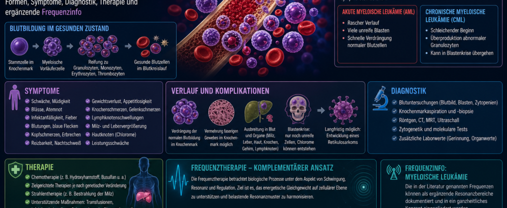
The Frequency therapy in myeloid leukemia In a complementary context, it is described as a supplementary perspective on the fundamentals of conventional medicine, hematopoiesis, bone marrow changes, symptoms, diagnosis, therapy, and possible resonance patterns. Myeloid leukemia is a malignant disease of the hematopoietic system in which cells of the myeloid lineage become malignant and can displace normal blood formation in the bone marrow.
From the perspective of conventional medicine, a distinction is primarily made between acute myeloid leukemia and chronic myeloid leukemia. Both forms affect the bone marrow but differ in terms of progression, cell maturity, course, and treatment. The literature also mentions certain resonance frequencies that are used as complementary therapies within the context of frequency therapy. Frequency info can be documented and examined.
Frequency Therapy for Myeloid Leukemia: An Overview from Conventional Medicine
Myeloid leukemia affects cells that normally develop in the bone marrow from immature precursor cells. These precursor cells give rise to, among other things, granulocytes, monocytes, red blood cells, and platelets. If one of these early cell lines becomes malignant, leukemia can develop.
In acute myeloid leukemia, immature myeloid cells—known as blasts—multiply rapidly. These cells do not mature normally and suppress healthy blood cell production. As a result, the disease can become life-threatening within a short period of time.
In chronic myeloid leukemia, a bone marrow cell undergoes a malignant transformation, producing large quantities of abnormal granulocytes. This disease may progress slowly at first, but can later enter a significantly more aggressive phase.
Frequency Therapy for Myeloid Leukemia and Blood Formation
The bone marrow is the central organ responsible for blood formation. This is where all the important blood cells are produced from stem cells. Red blood cells transport oxygen, white blood cells support the immune system, and platelets are essential for blood clotting.
In myeloid leukemia, this balance is disrupted. Leukemic cells accumulate in the bone marrow and displace the cells that produce normal blood cells. This results in characteristic changes in the blood count.
A lack of red blood cells leads to anemia. A low platelet count increases the risk of bleeding. A lack of functional white blood cells increases susceptibility to infection. At the same time, a large number of immature or abnormally altered white blood cells may appear in the blood.
Acute myeloid leukemia
Acute myeloid leukemia is a rapidly progressing, severe disease. In the literature, it is also described as myelotic, myelogenous, myeloblastic, or myelomonocytic leukemia. It is characterized by immature myeloid cells that become malignant and multiply rapidly.
These cells often quickly take over the normal function of the bone marrow. This leads to anemia, a tendency to bleed, and susceptibility to infections. Without treatment, acute myeloid leukemia can quickly become life-threatening.
The acute form can affect people of any age. It is more common in adults than in children, but can occur in people of all ages.
Chronic myeloid leukemia
Chronic myeloid leukemia is a disease in which a malignant bone marrow cell produces large quantities of abnormal granulocytes. These granulocytes can accumulate in the bone marrow, the blood, and various organs.
In the early stages, the disease may cause few symptoms for a long time. Some people do not learn they have the disease until they undergo a routine blood test. As the disease progresses, symptoms such as fatigue, weight loss, night sweats, fever, an enlarged spleen, and changes in blood counts may occur.
As the disease progresses, increasingly immature granulocytes may appear in the blood and bone marrow. If the disease continues to worsen, it can progress to what is known as a blast crisis. This phase resembles acute leukemia and is considered a serious turning point.
Frequency Therapy for Myeloid Leukemia and Bone Marrow Displacement
Leukemic cells spread throughout the bone marrow and displace normal cell lines. As a result, the bone marrow gradually loses its ability to produce healthy blood cells.
In addition, the literature describes how leukemic granulocytes can displace normal cells in the bone marrow and promote the formation of large amounts of fibrous tissue. This fibrous tissue can replace the normal bone marrow and further impair blood formation.
The bone marrow environment can change significantly, particularly in chronic cases. The balance between cell formation, cell maturation, and tissue structure is lost.
Spread of Leukemic Cells
Leukemia cells are not always confined to the bone marrow. They can enter the bloodstream and be carried to other organs, where they can continue to grow and divide.
The literature describes how leukemic cells can affect organs such as the spleen, liver, skin, bones, brain, lymph nodes, and other tissues. Small tumor masses may develop in or under the skin. Chloromas may also occur. These are tumor-like masses of leukemic cells.
If the central nervous system is affected, symptoms such as headaches, vomiting, irritability, or signs of meningitis may occur. Liver and kidney dysfunction may also develop as the disease progresses.
Blastic Crisis in Chronic Myeloid Leukemia
In chronic myeloid leukemia, further changes in the leukemic cells may occur as the disease progresses. If the abnormal stem cells suddenly begin to produce predominantly immature granulocytes, this is referred to as a blast crisis.
The blast crisis indicates that the disease has significantly worsened. The number of immature cells is rising sharply, while normal blood cells continue to be displaced.
During this phase, chloromas may develop in the skin, bones, brain, and lymph nodes. Symptoms often worsen rapidly. Anemia, a tendency to bleed, infections, fever, and organ involvement may become more severe.
Possible progression to reticulosarcoma
The literature describes how further dedifferentiation of myeloid leukemia cells over a prolonged period can lead to the development of a reticulosarcoma. This is a tumor-like lesion associated with highly degenerated cells.
This description illustrates that myeloid leukemias are not static. They can change over time, become more aggressive, and develop new biological characteristics.
For conventional medical evaluation, follow-up monitoring, blood tests, bone marrow findings, and cell maturation are therefore of great importance.
Common Symptoms of Myeloid Leukemia
The symptoms are primarily caused by impaired blood formation, organ involvement, and the proliferation of leukemic cells. They may appear rapidly in acute forms and develop gradually in chronic forms.
Possible complaints are
- Weakness
- Tiredness
- Paleness
- Shortness of breath
- Susceptibility to infection
- Fever
- Bleeding
- bruises
- Headache
- Vomiting
- Irritability
- Bone pain
- Joint pain
- Night sweats
- Weight loss
- Loss of appetite
- Swollen Lymph Nodes
- Enlarged spleen
- Skin nodules or chloromas
The symptoms are not always clear-cut. Chronic forms, in particular, may cause very few symptoms at first.
Acute symptoms caused by changes in blood counts
When the number of red blood cells is significantly reduced, anemia develops. This leads to paleness, rapid fatigue, shortness of breath, heart palpitations, and reduced performance.
A deficiency of blood platelets can cause signs of bleeding. These include nosebleeds, bleeding gums, pinpoint hemorrhages on the skin, bruises, or prolonged bleeding times.
A lack of functional white blood cells increases susceptibility to infection. This can lead to fever, recurrent infections, or severe cases of common infections.
Frequency Therapy for Myeloid Leukemia and Organ Involvement
Leukemia cells can accumulate in organs. The spleen and liver, in particular, may be enlarged. An enlarged spleen can cause a feeling of pressure or pain in the upper left abdomen. Some patients report feeling full early because the enlarged spleen presses on the stomach.
Lymph nodes may swell. Skin nodules may develop when leukemic cells form clusters in the skin or subcutaneous tissue.
If the central nervous system is affected, headaches, vomiting, irritability, neurological abnormalities, or signs of meningeal irritation may occur. Bone and joint pain may also result from bone marrow involvement or leukemic infiltration.
Conventional Medical Diagnosis of Myeloid Leukemia
Diagnosis usually begins with blood tests. The complete blood count reveals changes in white blood cells, red blood cells, and platelets. Often, immature cells, elevated or decreased cell counts, and signs of impaired blood formation are observed.
A bone marrow biopsy or bone marrow aspiration is essential for confirming the diagnosis. These procedures are used to determine which cell lineages are proliferating, what percentage of immature blasts is present, and what maturation abnormalities exist.
In addition, imaging techniques such as X-rays, computed tomography, magnetic resonance imaging, and ultrasound may be used. These tests help assess organ enlargement, bone damage, lymph node involvement, or other abnormalities.
Additional Diagnostic Considerations
In addition to blood tests and bone marrow examinations, cytogenetic and molecular tests are important today. They help identify genetic changes that can be critical for diagnosis, prognosis, and treatment planning.
In chronic myeloid leukemia, the detection of typical genetic alterations plays a central role. In acute myeloid leukemia, various subgroups are distinguished based on genetic and cellular characteristics.
Organ function, coagulation parameters, infection markers, and metabolic values are also frequently monitored, as the disease can affect the entire body.
Conventional Medical Treatment for Myeloid Leukemia
Treatment depends on whether the disease is acute or chronic, the patient’s genetic profile, age, general health, and the stage of the disease. The literature mentions chemotherapy, hydroxyurea, busulfan, and radiation therapy to the spleen.
In cases of acute myeloid leukemia, intensive chemotherapy is often the primary treatment. The goal is to suppress leukemic cells and restore normal blood production.
In chronic myeloid leukemia, treatment depends on the stage of the disease and the molecular findings. In addition, measures may be necessary to control high cell counts, treat splenomegaly, and support blood cell production.
Supportive Care for Myeloid Leukemia
In addition to directly treating the leukemia cells, supportive care is important. This may include blood transfusions, platelet transfusions, treatment of infections, fever reduction, pain management, and protective measures for patients with immunodeficiency.
Since normal blood production is impaired, blood tests must be monitored regularly. Infections can be severe and require prompt attention.
Nutrition, physical rest, psychological stabilization, and close medical monitoring also play an important role in the overall course of the disease.
Frequency Therapy for Myeloid Leukemia in a Complementary Context
Frequency therapy looks at biological processes from the point of view of vibration, Resonance and regulation. In myeloid leukemia, the complementary approach focuses on the bone marrow, hematopoiesis, immature cell lineages, the immune status, organ involvement, possible microbial resonance patterns, and systemic stress.
The literature cites certain resonance frequencies that are described in connection with myeloid leukemia. These frequencies can be documented as supplementary frequency information in the context of frequency therapy.
The focus is not only on the individual frequency, but on the overall resonance pattern: bone marrow, blood cells, the immune system, the spleen, the liver, the lymphatic system, and any potential accompanying stressors are all considered together.
A Broader Look at Frequency Therapy and Leukemia
Leukemias are diseases of the entire hematopoietic system. They do not affect just a single organ, but impact the blood, bone marrow, immune system, metabolism, organs, and often overall physical performance as well.
Frequency therapy views such conditions, in a broader sense, as manifestations of disrupted biological order and altered Cell communication. In myeloid leukemia, the primary features are the impaired maturation of myeloid cells, the suppression of normal hematopoiesis, and the systemic spread of leukemic cells.
The frequencies cited in the literature can serve as a complementary guide for documenting resonance patterns and integrating them into a holistic framework.
Prevalence Information: Myeloid Leukemia
The following frequencies are cited in the literature in connection with myeloid leukemia. In the complementary context of frequency therapy, they are regarded as supplementary resonance ranges.
Frequency therapy for myeloid leukemia
311 kHz,
314 kHz,
330–340 kHz,
353 kHz,
372 kHz,
402-410 kHz,
420–437 kHz,
446 kHz,
450–452 kHz,
461–469 kHz,
496 kHz,
513–515 kHz,
540–544 kHz,
558–559 kHz,
567–573 kHz.
These frequencies are described in the literature as common resonance frequencies associated with myeloid leukemia. In the context of frequency therapy, they can be used as a supplementary guide for documentation, resonance analysis, and individualized treatment.
Frequency information: mid-range frequencies
Some of the frequencies lie in the midrange and can be documented as a central resonance group in the complementary frequency analysis.
Frequency therapy for moderate resonances
311 kHz,
314 kHz,
330–340 kHz,
353 kHz,
372 kHz,
402-410 kHz,
420–437 kHz.
These frequency ranges can be considered in relation to bone marrow, the myeloid cell line, the immune status, and the leukemia process.
Frequency information: higher resonance ranges
In addition to the mid-frequency ranges, the literature mentions several higher resonance ranges.
Frequency therapy for higher resonances
446 kHz,
450–452 kHz,
461–469 kHz,
496 kHz,
513–515 kHz,
540–544 kHz,
558–559 kHz,
567–573 kHz.
These higher frequencies complement the resonance pattern observed in myeloid leukemia. In particular, the frequency ranges 461–469 kHz, 540–544 kHz, and 567–573 kHz can be documented as additional resonance fields in frequency therapy.
Frequency Information: Note on Completeness
The literature indicates that the frequency list provided may be incomplete. In complementary frequency therapy, this means that the listed frequencies should be viewed as a documented guide, while individual resonance patterns may also be taken into account.
Frequency therapy tailored to individual resonance frequencies
Bone marrow,
Blood formation,
myeloid cell line,
Blasts,
Granulocytes,
Spleen,
Liver,
Lymphatic system,
individual resonance patterns.
This additional perspective can help us view the frequency lists not in a rigid manner, but in the context of each individual situation.
Frequency Therapy for Myeloid Leukemia: A Comparison of Frequency Patterns
When comparing the frequencies, several broad bands stand out. Particularly noticeable are 330–340 kHz, 402–410 kHz, 420–437 kHz, 461–469 kHz, 540–544 kHz, and 567–573 kHz.
Individual frequencies such as 311 kHz, 314 kHz, 353 kHz, 372 kHz, 446 kHz, and 496 kHz complement these broader frequency fields. In complementary frequency therapy, this combination can be understood as a multilayered resonance pattern.
The frequency lists are always considered in conjunction with the conventional medical diagnosis, whether the condition is acute or chronic, the blood count, the bone marrow findings, organ involvement, and the individual’s regulatory status.
Frequency Therapy for Myeloid Leukemia: Summary
Myeloid leukemia is a malignant disease of the hematopoietic system. In the acute form, immature myeloid cells multiply rapidly and displace normal blood cell production. In the chronic form, large numbers of abnormal granulocytes are produced, which can accumulate in the bone marrow, blood, and organs.
Typical symptoms include fatigue, weakness, pallor, shortness of breath, infections, fever, bleeding, bone pain, joint pain, night sweats, weight loss, swollen lymph nodes, and an enlarged spleen. In advanced stages, blast crises, chloromas, and organ involvement may occur.
Frequency therapy offers a complementary perspective. The literature cites resonance frequencies such as 311 kHz, 314 kHz, 330–340 kHz, 353 kHz, 372 kHz, 402–410 kHz, 420–437 kHz, 446 kHz, 450–452 kHz, 461–469 kHz, 496 kHz, 513–515 kHz, 540–544 kHz, 558–559 kHz, and 567–573 kHz. These frequency lists can be used in a complementary context for documentation, resonance analysis, and individualized frequency therapy work.



Comments are closed, but trackbacks and pingbacks are open.