





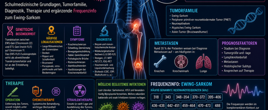
The Frequency therapy in Ewing's sarcoma is described in the complementary context as a supplementary perspective on the fundamentals of conventional medicine, tumor development, symptoms, diagnosis, treatment, and possible resonance patterns. Ewing sarcoma is a highly malignant Tumor, which primarily affects bones and soft tissues. It belongs to a family of tumors that share common biological and genetic characteristics.
From a conventional medical perspective, Ewing sarcoma is classified as an aggressive form of cancer that primarily affects children, adolescents, and young adults. It often develops in long bones, the pelvis, the ribs, or the area around the shoulder blade. The literature also mentions certain resonance frequencies that are used as complementary therapies in the context of frequency therapy. Frequency info can be documented and examined.
Frequency Therapy for Ewing Sarcoma: An Overview from Conventional Medicine
Ewing sarcoma is a malignant primary bone tumor. It is often associated with red bone marrow and shares histological similarities with small round cell tumors. Although it is classically described as a bone tumor, it can also occur outside the bone in soft tissues.
The Ewing sarcoma family includes several closely related tumor types. These include classic Ewing sarcoma, peripheral primitive neuroectodermal tumor, neuroepithelioma, atypical Ewing sarcoma, and Askin tumor of the chest wall.
These tumors are grouped together based on their common characteristics. The literature suggests that they may originate from cells associated with the neural crest or certain neuronal progenitor cells. This classification indicates that they are not merely a local bone tumor but rather a biologically complex group of tumors.
Frequency Therapy for Ewing's Sarcoma and Related Tumors
Tumors in the Ewing sarcoma family share similar cellular and genetic characteristics. They can occur in bones or soft tissues and, depending on their location, can cause a wide variety of symptoms.
The peripheral primitive neuroectodermal tumor, or PNET for short, exhibits a higher degree of neuroectodermal differentiation. Neuroepithelioma is also associated with nerve-like cellular features. Askin’s tumor typically affects the chest wall and may present with pain, breathing difficulties, or mass effect.
Atypical Ewing sarcoma is described as a variant within this family of tumors. Accurate histological and molecular genetic evaluation is crucial, as these tumors are similar in terms of treatment but may have different courses depending on their extent and location.
Genetic Characteristics of Ewing Sarcoma
In modern conventional medical thinking, a specific genetic mutation plays a central role. The literature describes that tumors of the Ewing sarcoma family frequently exhibit a translocation. In this process, the EWS gene on chromosome 22 is fused with a gene from the ETS family on chromosome 11.
An EWS-FLI1 fusion protein is formed particularly frequently in this process. This fusion protein can act as a misdirected transcription factor. This means that it alters the activity of certain genes and can thereby influence growth, differentiation, and cell behavior.
This genetic characteristic is significant for the development and biological activity of the tumors. It also explains why molecular diagnostics play an important role in Ewing sarcoma.
Common Sites of Ewing Sarcoma
Ewing sarcoma often occurs in the long bones of the extremities. The femur, tibia, humerus, and other long bones are particularly susceptible. The tumor can develop both in the shaft of the bone and in the transitional zones.
The pelvis is also a common site. Tumors in the pelvic region can go unnoticed for a long time because they are initially deep-seated and do not cause pain or functional impairment until later. In addition, the ribs, shoulder blade, and chest wall are described as possible sites of origin.
Paraspinal, retroperitoneal, or deep pelvic tumors can cause back pain or vague deep pain. Depending on their location, they can also affect nerves, blood vessels, organs, or motor function.
Frequency Therapy for Ewing's Sarcoma and Metastasis
The prognosis depends largely on whether the disease is localized at the time of diagnosis or has already Metastases has developed. The literature describes that many patients initially have localized disease, while some already have metastatic tumor foci.
Metastases occur particularly frequently in the bones, bone marrow, and lungs. These sites are therefore especially important in assessing the extent of the disease.
If the bone marrow is affected, changes in the blood count may occur. In cases of severe bone marrow involvement, signs of bleeding such as petechiae or purpura may occur, as the platelet count may be reduced. In cases of lung metastases, pleural signs, shortness of breath, or asymmetric breath sounds may occur.
Possible Symptoms of Ewing Sarcoma
Symptoms depend heavily on the location of the tumor. The disease often begins with pain in the affected bone or soft tissue area. This pain may initially seem to be related to physical activity but may later become constant.
Possible symptoms are
- localized pain
- Swelling
- Overheating
- Limited mobility
- Pain on pressure
- Fever
- Weight loss
- general weakness
- Back Pain Caused by Deep-Seated Tumors
- pathological fractures
- Breathing difficulties caused by lung or chest wall involvement
- Signs of Bleeding in Cases of Bone Marrow Involvement
Fever and weight loss may indicate advanced or metastatic disease. Pathological fractures can occur when the tumor weakens the stability of a bone.
Frequency Therapy for Ewing Sarcoma and Differential Diagnosis
Ewing sarcoma can cause symptoms that initially resemble those of inflammation, infection, a sports injury, or other bone disorders. Pain, swelling, and fever, in particular, can make it difficult to distinguish the condition from these other conditions.
From a conventional medical perspective, it is therefore important to rule out other conditions. This may include blood tests, such as markers of inflammation, a complete blood count, blood cultures, and other laboratory parameters. These tests help to better identify infectious processes, inflammatory diseases, or other causes.
However, a definitive diagnosis is hindered by Biopsy, histological examination, and immunohistochemical analysis. In addition, molecular genetic testing may provide evidence of typical fusion genes.
Conventional Medical Diagnosis of Ewing Sarcoma
The diagnostic process involves several steps. First, symptoms, physical findings, and imaging studies are evaluated. X-rays can provide evidence of bone changes. Computed tomography and magnetic resonance imaging help to more accurately visualize the extent of the condition in the bone and soft tissues.
Positron emission tomography (PET) or FDG-PET can be used to detect metabolically active tumor foci and assess their spread throughout the body. Screening for lung metastases and bone marrow involvement also plays an important role.
The biopsy is crucial. Only by examining the tumor tissue can a definitive diagnosis be made. Immunohistochemistry and molecular analyses can confirm that the tumor belongs to the Ewing sarcoma family.
Conventional Medical Treatment of Ewing's Sarcoma
Treatment for Ewing sarcoma is usually multimodal. This means that several treatment methods are combined. The literature mentions chemotherapy, radiation therapy, and specific surgical procedures.
Chemotherapy is a key component of treatment, as Ewing sarcoma is considered a systemic disease. Even if the tumor appears to be localized, microscopic tumor metastases may be present. For this reason, drug therapy is usually initiated early on.
The surgical procedure depends on the location, size, and extent of the tumor. The goal is to remove as much of the tumor as possible, to the extent that this is functionally and anatomically feasible. Radiation therapy may be used as an adjunct, particularly when complete surgical removal is difficult or certain risk factors are present.
Prognostic Factors in Ewing Sarcoma
The prognosis depends heavily on the stage of the disease at the time of diagnosis. Patients with localized disease have a better prognosis than those with metastases. Involvement of the bone marrow, multiple bone regions, or the lungs can be particularly unfavorable.
Other factors include tumor size, tumor location, response to chemotherapy, success of surgical removal, and the tumor's biological characteristics. The type of genetic alteration may also be relevant for research and prognosis assessment.
Rapid and accurate diagnosis is therefore crucial for planning early and targeted treatment.
Frequency Therapy for Ewing Sarcoma and Possible Associated Patterns
In the literature, the development of Ewing sarcoma is associated with a genetic predisposition and a combined infectious burden. A sarcoma virus and human lymphotropic viruses are mentioned Viruses and especially often Mycoplasma fermentans. In addition, numerous other secondary infections are mentioned.
In complementary frequency-based thinking, such data are viewed as potential resonance patterns. In this approach, the tumor, immune status, bone marrow, tissue environment, and associated microbial burdens are not considered in isolation from one another, but are integrated into a holistic picture.
In this context, frequency therapy can serve as a complementary perspective. It identifies resonance ranges that are mentioned in the literature in connection with Ewing sarcoma.
Frequency Therapy for Ewing's Sarcoma in a Complementary Context
Frequency therapy looks at biological processes from the point of view of vibration, Resonance and regulation. In Ewing sarcoma, the complementary focus is on bone, bone marrow, soft tissue, immune responses, possible viral and mycoplasmal infections, and the tendency for systemic spread.
The literature cites specific resonance frequencies associated with Ewing sarcoma. These frequencies can be used as supplementary frequency information in the context of frequency therapy.
The focus here is on documentation: Which frequency ranges are mentioned? What overlaps exist with other infectious or neoplastic resonance fields? How can this information be integrated into a holistic approach?
Frequency therapy and cancer in an expanded view
Cancers such as Ewing's sarcoma do not result solely from localized cell growth. They affect Cell communication, gene regulation, tissue microenvironment, immune response, bone marrow, and possible systemic stress patterns.
In Ewing sarcoma, the genetic fusion mutation is a key characteristic. At the same time, the literature also mentions accompanying infectious factors. From the perspective of frequency therapy, both levels can be considered together: structural cellular changes and a resonance-based analysis of the microenvironment.
A clear conventional medical diagnosis and treatment plan are crucial, especially in cases of aggressive sarcomas. The supplementary frequency information can serve as an additional tool for complementary documentation and individual resonance analysis.
Frequency Information: Ewing Sarcoma
The following frequencies are cited in the literature in connection with Ewing sarcoma. In the complementary context of frequency therapy, they are regarded as supplementary resonance ranges.
Frequency Therapy for Ewing's Sarcoma
318 kHz,
343 kHz,
348–354 kHz,
370–372 kHz,
395–406 kHz,
436–438 kHz,
442-451 kHz,
459-464 kHz,
470–473 kHz,
488 kHz,
491–493 kHz,
496 kHz,
511–519 kHz,
523–527 kHz,
530–540 kHz.
These frequencies are described in the literature as common resonances associated with Ewing sarcoma. In the context of frequency therapy, they can be used as a supplementary guide for documentation, resonance analysis, and individualized treatment.
Frequency information: mid-range frequencies
Some of the frequencies fall within the midrange and form a distinctive group that deserves special attention in the complementary analysis.
Frequency therapy for moderate resonances
318 kHz,
343 kHz,
348–354 kHz,
370–372 kHz,
395–406 kHz,
436–438 kHz,
442–451 kHz.
In frequency therapy, these ranges can be documented as key resonance fields in Ewing sarcoma. In particular, the 395–406 kHz and 442–451 kHz ranges appear as broad frequency fields that may be significant in a complementary approach.
Frequency information: higher resonance ranges
In addition to the mid-range resonances, the literature mentions several higher-frequency bands.
Frequency therapy for higher resonances
459-464 kHz,
470–473 kHz,
488 kHz,
491–493 kHz,
496 kHz,
511–519 kHz,
523–527 kHz,
530–540 kHz.
These frequency ranges can be considered additional resonance fields. They complement the mid-range frequencies and, together, form a comprehensive list of frequencies associated with Ewing sarcoma.
Frequency Information: Possible Microbial Resonance Patterns
In the literature, Ewing sarcoma is associated with a sarcoma virus, human lymphotropic viruses, and—particularly frequently—Mycoplasma fermentans as possible patterns of infection. Other secondary infections may also occur.
Frequency Therapy for Concomitant Conditions
Sarcoma virus,
human lymphotropic viruses,
Mycoplasma fermentans,
secondary bacterial and viral infections.
These accompanying patterns are not considered in isolation within the complementary context. They are incorporated into the frequency analysis along with tumor location, bone marrow, the immune system, the surrounding tissue, and individual regulatory capacity.
Frequency Therapy for Ewing Sarcoma: Comparison of Frequency Patterns
When comparing the frequencies, a broad distribution across several resonance bands is evident. Particularly noticeable are the ranges 348–354 kHz, 395–406 kHz, 442–451 kHz, 511–519 kHz, 523–527 kHz, and 530–540 kHz.
In frequency therapy, this gradation can be interpreted as an indication of multiple resonance levels. Individual frequencies such as 318 kHz, 343 kHz, 488 kHz, and 496 kHz complement the larger frequency blocks.
The frequency lists are always considered in conjunction with the conventional medical diagnosis, the type of tumor, its spread, its location, and the patient’s individual regulatory status.
Frequency Therapy for Ewing Sarcoma: Summary
Ewing sarcoma is a highly malignant tumor of the bone and soft tissue regions. It belongs to a family of tumors that also includes peripheral primitive neuroectodermal tumors, neuroepitheliomas, atypical Ewing sarcomas, and Askin tumors.
In conventional medicine, genetic alterations such as the EWS-FLI1 fusion protein, tumor location, metastasis, bone marrow involvement, and lung involvement are of great importance. Diagnosis is made through imaging, biopsy, immunohistochemical examination, molecular analysis, and staging. Treatment usually consists of chemotherapy, surgery, and radiation therapy.
Frequency therapy offers a complementary perspective. In the literature, resonance frequencies such as 318 kHz, 343 kHz, 348–354 kHz, 370–372 kHz, 395–406 kHz, 436–438 kHz, 442–451 kHz, 459–464 kHz, 470–473 kHz, 488 kHz, 491–493 kHz, 496 kHz, 511–519 kHz, 523–527 kHz, and 530–540 kHz. These frequency lists can be used in a complementary context for documentation, resonance analysis, and individualized frequency therapy work.

Comments are closed, but trackbacks and pingbacks are open.