





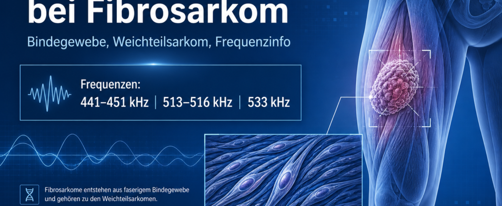
The Frequency therapy in fibrosarcoma is described in the complementary context as a supplementary perspective on the fundamentals of conventional medicine, symptoms, diagnosis, treatment, and possible resonance patterns. Fibrosarcoma is a malignant Tumor, which originates from fibrous connective tissue. It is characterized by immature fibroblasts or dedifferentiated spindle-shaped tumor cells.
From a conventional medical perspective, fibrosarcoma is classified as a sarcoma. It can present as a soft-tissue tumor or develop as a primary or secondary bone tumor. The literature also mentions specific resonance frequencies that are used as a complementary treatment in the context of frequency therapy Frequency info can be documented and examined.
Frequency Therapy for Fibrosarcoma: An Overview from Conventional Medicine
Fibrosarcoma is a malignant tumor of the connective tissue. It arises from fibroblastic cells—that is, cells that are normally involved in the formation of connective tissue fibers and collagen. When these cells become malignant, they can grow uncontrollably and displace or destroy surrounding tissue.
Histologically, fibrosarcoma often shows immature, rapidly proliferating fibroblasts or anaplastic spindle cells. The term „anaplastic“ describes cells that have largely lost their normal differentiation and exhibit distinct malignant features.
Fibrosarcomas can exhibit varying degrees of malignancy. There are low-grade tumors, intermediate-grade tumors, and highly malignant, anaplastic variants. The tumor grade is particularly important for prognosis, treatment, and the risk of recurrence.
Frequency Therapy for Fibrosarcoma and Tumor Development
Fibrous connective tissue is present throughout the body. For this reason, a fibrosarcoma can develop in various locations. Soft tissues, areas surrounding muscles, fascial regions, and bone structures are particularly susceptible.
A fibrosarcoma can present as a soft-tissue mass. The tumor originates in the connective tissue of the soft tissues and often grows deep beneath the surface. Such tumors can go unnoticed for a long time because they cause little or no pain at first.
In addition, a fibrosarcoma can occur as a bone tumor. A distinction is made between primary and secondary fibrosarcomas of the bone. This classification is important for medical evaluation because their origin, behavior, and prognosis may differ.
Primary Fibrosarcoma of the Bone
Primary osteofibrosarcoma originates directly in the bone. It is a malignant fibroblastic tumor that can produce varying amounts of collagen. Collagen is an important structural protein of connective tissue.
A primary fibrosarcoma can arise centrally within the medullary cavity of the bone. The medullary cavity is the area that contains bone marrow. Alternatively, the tumor can also arise peripherally from the periosteum. This layer is called the periosteum and surrounds the bone from the outside.
Depending on where it originates, the tumor can weaken the bone from the inside or invade the bone structure from the outside. This can compromise the bone's stability.
Secondary Fibrosarcoma of the Bone
Secondary fibrosarcoma of the bone arises from a preexisting lesion or following prior radiation therapy to bone or soft tissue. In the literature, this form is described as particularly aggressive.
Secondary fibrosarcomas can develop from preexisting lesions. In rare cases, a malignant tumor may also develop later following radiation therapy to a specific area of the body. Such radiation-associated sarcomas are considered a serious late complication.
The prognosis for secondary fibrosarcomas is described in the literature as poor, especially if the tumor is high-grade, large, or already advanced.
Common Sites of Fibrosarcoma
Fibrosarcomas can occur in many parts of the body. The literature specifically mentions areas around the knee, thigh, arm, and hip. Deep soft tissues and muscular fasciae may also be affected.
Soft-tissue fibrosarcomas often develop deep within the muscle fascia. As a result, they can grow to a relatively large size before they are noticed. Visible swelling often does not appear until a late stage.
Fibrosarcomas near the bone can cause pain, swelling, and, later on, structural weakening. If the bone is severely affected, a pathological fracture may occur. In this case, the bone does not break as a result of a normal accident, but because its stability has been compromised by the tumor.
Symptoms of Fibrosarcoma
Symptoms depend on whether the tumor originates in the bone or in the soft tissues. Bone sarcomas often do not cause pain or swelling until some time has passed. Initially, the symptoms may be nonspecific and resemble those of overexertion, inflammation, or injury.
Possible symptoms of bone involvement include:
- increasing pain
- local swelling
- Pressure sensitivity
- Stress-induced pain
- Rest pain in later stages
- Limited mobility
- Bone Instability
- pathological fracture
Soft-tissue fibrosarcomas are often initially painless space-occupying lesions. They appear as firm, minimally movable masses. The affected area may be tender to the touch, but it does not necessarily have to be painful.
Advanced Symptoms of Fibrosarcoma
As a fibrosarcoma grows, it can displace or compress surrounding structures. In advanced cases, nerves or blood vessels may be affected. This can lead to neurological or vascular changes.
Possible advanced symptoms include:
- Numbness
- Tingling
- Nerve pain
- Muscle weakness
- Circulatory Disorders
- Swelling caused by venous congestion
- limited mobility
- Loss of function in the affected region
Deep-seated tumors can grow for a long time before causing symptoms. Therefore, any firm, enlarging, or deep mass always requires further evaluation.
Frequency Therapy for Fibrosarcoma and Diagnosis
Conventional medical diagnosis begins with a clinical examination and imaging. Computed tomography, magnetic resonance imaging, and positron emission tomography can be used to assess the location, size, extent, and possible spread of the tumor.
Magnetic resonance imaging is particularly important for visualizing soft tissues, muscles, fascia, nerves, and blood vessels. Computed tomography can show bone changes more accurately. A PET scan can help identify metabolically active tumor regions or possible Metastases to record.
The final diagnosis is made by Biopsy and histological examination. This involves removing a tissue sample and analyzing it under a microscope. Special staining techniques can help distinguish fibrosarcoma from other tumors.
Differential Diagnosis of Fibrosarcoma
Fibrosarcoma must be distinguished from other bone and soft tissue tumors. The differential diagnosis is important because different types of tumors require different treatments and have different prognoses.
The literature cites osteosarcoma, Paget's sarcoma, malignant fibrous histiocytoma, fibrous dysplasia, and fibrous histiocytoma, among others, as important differential diagnoses.
Inflammatory changes, benign soft-tissue tumors, scar tissue, hematomas, or other sarcomas can also appear similar. Therefore, a combination of imaging, biopsy, histology, and special stains is crucial.
Conventional Medical Treatment of Fibrosarcoma
Treatment depends on the tumor grade, size, location, extent of spread, operability, and the patient's overall condition. A combination of surgery, radiation therapy, and chemotherapy is often used.
The goal of the surgery is to remove the tumor as completely as possible. The aim is to ensure a sufficient safety margin to reduce the risk of recurrence. Particularly in the case of tumors on the arms, legs, or in the pelvic area, the function of the affected region must also be taken into account.
Radiation therapy can be used to improve local tumor control. Chemotherapy can help reduce the risk of metastasis or treat advanced cancer systemically. The specific treatment strategy is determined on an individual basis.
Frequency Therapy for Fibrosarcoma in a Complementary Context
Frequency therapy looks at biological processes from the point of view of vibration, Resonance and regulation. In the case of fibrosarcoma, the complementary focus is on connective tissue, fibroblasts, collagen formation, the tissue microenvironment, tumor growth, and possible infectious resonance patterns.
The literature describes a causal link to viruses. The frequency ranges provided are considered supplementary frequency information in the context of frequency therapy. They can be used for documentation, resonance analysis, and as a guide for individualized frequency therapy.
Frequency therapy is integrated into a holistic approach. The tumor is not viewed merely as a localized mass of tissue, but is also considered in relation to connective tissue structure, immune status, cell regulation, and potential systemic stress.
Frequency therapy and cancer in an expanded view
Cancers such as fibrosarcoma arise from changes in cell growth, differentiation, tissue architecture, and biological communication. In fibrosarcoma, these changes particularly affect the cells that are normally involved in the formation of fibrous connective tissue.
Frequency therapy views cancer, in a broader sense, as a manifestation of disrupted order and altered resonance within the body. In the case of sarcomas, factors such as the tissue microenvironment, connective tissue regulation, susceptibility to inflammation, immune response, and potential pathogenic patterns may be taken into account.
An accurate conventional medical diagnosis and treatment are crucial, especially in cases of deep-seated or aggressive sarcomas. The complementary frequency information can also be used as a documentary and complementary perspective.
Frequency Information: Fibrosarcoma
The following frequencies are cited in the literature in connection with fibrosarcoma. In the complementary context of frequency therapy, they are regarded as supplementary resonance ranges.
Frequency Therapy for Fibrosarcoma
441–451 kHz,
513–516 kHz,
533 kHz.
These frequencies are referred to in the literature as common resonances of the described Virus mentioned in connection with fibrosarcoma. In the context of frequency therapy, they can be used as a supplementary guide for documentation, resonance analysis, and individualized work.
Frequency information: mid-range frequencies
In the literature, the 441–451 kHz range is considered the central mid-frequency resonance band for fibrosarcoma.
Frequency therapy for moderate resonances
441–451 kHz.
This frequency range deserves special attention in the complementary context, as it is described as a broad resonance field. It can be used in frequency therapy to document possible virus-related stress patterns.
Frequency information: higher resonance ranges
In addition, higher resonances are mentioned that complement the frequency profile in fibrosarcoma.
Frequency therapy for higher resonances
513–516 kHz,
533 kHz.
These frequencies can be considered as additional resonance points in the complementary frequency analysis. Together with 441–451 kHz, they form the list of frequencies associated with fibrosarcoma as cited in the literature.
Frequency Therapy for Fibrosarcoma: Comparison of Frequency Patterns
For fibrosarcoma, the frequency list is relatively compact. It covers a broad mid-range resonance band of 441–451 kHz, as well as higher ranges at 513–516 kHz and 533 kHz.
In frequency therapy, this structure can be understood as a clearly defined resonance pattern. The 441–451 kHz range is the primary focus, while 513–516 kHz and 533 kHz represent supplementary higher resonance points.
These frequencies are always considered in conjunction with the conventional medical diagnosis, the tumor grade, the location, the extent of the disease, and the individual’s regulatory status.
Frequency Therapy for Fibrosarcoma: Summary
Fibrosarcoma is a malignant tumor of fibrous connective tissue. It can occur as a soft-tissue sarcoma or as a primary or secondary bone tumor. It is characterized by immature fibroblasts, anaplastic spindle cells, and, depending on the type, varying degrees of aggressive growth.
In conventional medicine, the focus is on imaging, biopsy, histological examination, and differential diagnosis. Differential diagnoses include, among others, osteosarcoma, Paget’s sarcoma, malignant fibrous histiocytoma, fibrous dysplasia, and fibrous histiocytoma. Treatment may include surgery, radiation therapy, chemotherapy, and symptomatic measures.
Frequency therapy offers a complementary perspective. The literature cites resonance frequencies of 441–451 kHz, 513–516 kHz, and 533 kHz for fibrosarcoma. These frequency lists can be used in a complementary context for documentation, resonance analysis, and individualized frequency therapy work.

Comments are closed, but trackbacks and pingbacks are open.