





The Frequency therapy in rhabdomyosarcoma is described in the complementary context as a supplementary perspective on the fundamentals of conventional medicine, tumor types, diagnostics, therapy, and possible resonance patterns. Rhabdomyosarcoma is a malignant Tumor, which can develop from immature precursor cells of the skeletal muscles. Although this tumor is associated with muscle tissue, it can occur in a wide variety of locations throughout the body.
From the perspective of conventional medicine, rhabdomyosarcoma is classified as a soft-tissue sarcoma. It is particularly significant in childhood, as it is one of the most common malignant soft-tissue tumors in children. The literature also describes certain resonance frequencies that are used as a complementary therapy in the context of frequency therapy Frequency info can be documented and examined.
Frequency Therapy for Rhabdomyosarcoma: An Overview of Conventional Medicine
Rhabdomyosarcoma is a malignant tumor whose cells exhibit characteristics of immature muscle progenitor cells. These cells originate from primitive mesenchymal tissue and have the ability to develop into skeletal muscle. For this reason, rhabdomyosarcoma is described as a tumor with striated muscle differentiation.
The tumor may occur near muscle tissue, but it does not necessarily have to originate directly from a developed muscle. It can occur in many parts of the body, such as the head and neck region, the urogenital tract, the extremities, the trunk, the abdomen, the chest, the digestive tract, and the area around the anus and perineum.
Rhabdomyosarcoma is particularly common in children. In the head and neck region, it primarily affects children under the age of ten. There, it is one of the most common malignant soft-tissue tumors.
Frequency Therapy for Rhabdomyosarcoma and Typical Locations
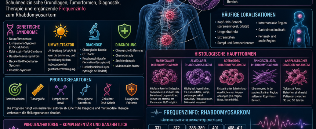
Rhabdomyosarcomas can develop almost anywhere in the body, with the exception of the bone itself. Tumors in the head and neck region are particularly common. Within this region, the orbit and parameningeal areas are of particular importance. Parameningeal means that the tumor is located near the meninges or near anatomically sensitive structures of the head.
Other common sites include the urogenital tract, the extremities, the trunk, the posterior abdomen, the chest, the gastrointestinal tract, and the perianal and anal regions.
The location of the tumor is critical for prognosis and treatment planning. Tumors in easily accessible locations are often easier to remove surgically than tumors in deep, complex, or sensitive areas.
Rhabdomyosarcoma in Children
Rhabdomyosarcoma is of particular importance in pediatric oncology. It can occur very early in life and follows different courses depending on the subtype, location, and extent of the disease.
Children with rhabdomyosarcoma require particularly careful diagnosis, as tumors in growing bodies can have complex effects. In addition to tumor size and spread, function, organ preservation, and future development are also important aspects of treatment planning.
Tumors in the head and neck region can affect the eyes, sinuses, base of the skull, oral cavity, or pharynx. In the urogenital tract, the bladder, prostate, testicular region, vagina, or other pelvic structures may be involved.
Frequency Therapy for Rhabdomyosarcoma and Genetic Syndromes
The literature describes various genetic syndromes that may be associated with an increased incidence of rhabdomyosarcomas. These include neurofibromatosis, Li-Fraumeni syndrome, Rubinstein-Taybi syndrome, basal cell nevus syndrome, Beckwith-Wiedemann syndrome, and Costello syndrome.
Li-Fraumeni syndrome is associated with mutations in the TP53 tumor suppressor gene. This gene plays a central role in regulating cell growth, DNA damage, and tumor suppression. When this regulatory function is disrupted, the risk of developing various types of cancer may be increased.
Other genetic syndromes also indicate that rhabdomyosarcomas are not merely isolated local tumors, but may be linked to deeper regulatory and developmental mechanisms.
Major histological types of rhabdomyosarcoma
In conventional medicine, rhabdomyosarcoma is classified into several major histological subtypes. These subtypes differ in terms of age, location, cellular appearance, genetic characteristics, and biological behavior.
The literature describes five major groups:
- embryonal rhabdomyosarcoma
- alveolar rhabdomyosarcoma
- embryonal botryoides rhabdomyosarcoma
- spindle-cell embryonal rhabdomyosarcoma
- anaplastic rhabdomyosarcoma
Accurate histological classification is of great importance for prognosis and treatment.
Frequency therapy for embryonal rhabdomyosarcoma
Embryonal rhabdomyosarcoma is the most common subtype in children. It occurs particularly frequently in the head and neck region and in the urogenital tract.
Under the microscope, this type of tumor exhibits great cellular diversity. This means that the cells may reflect different stages of skeletal muscle development. This diversity is consistent with the idea that the tumor arises from immature mesenchymal cells that have retained the ability to differentiate into muscle.
In the literature, embryonal rhabdomyosarcoma is associated with the loss of certain genetic Information described on the short arm of chromosome 11. The 11p15 region is specifically mentioned. This loss may indicate the presence of a tumor suppressor region in that location, the disruption of which could play a significant role in tumor development.
Frequency Therapy for Alveolar Rhabdomyosarcoma
Alveolar rhabdomyosarcoma is more commonly reported in adolescents. It primarily affects the extremities, the trunk, and the perianal and perirectal regions.
This subtype often exhibits characteristic genetic alterations. The literature mentions PAX3 and PAX7 translocations. Individuals with a PAX7 translocation are often younger and may have a longer event-free survival time than those with a PAX3 translocation.
Unlike embryonal rhabdomyosarcoma, alveolar rhabdomyosarcoma frequently exhibits gene amplifications. The DNA content is typically described as tetraploid. These biological characteristics may indicate a different growth and risk profile.
Botryoides Rhabdomyosarcoma
Botryoid rhabdomyosarcoma is a specific form of embryonic rhabdomyosarcoma. It typically arises beneath the mucosal surfaces of body orifices.
Common sites include the vagina, the bladder, and the nasal cavity. The term „botryoid“ refers to a grape-like growth pattern. Such tumors may appear polypoid and protrude into cavities.
Precisely because these tumors develop on mucous membranes, they may present with symptoms such as bleeding, discharge, urinary symptoms, or visible masses.
Spindle-cell rhabdomyosarcoma
Spindle-cell rhabdomyosarcoma is described in the literature primarily in the paratesticular region. This region is located in the area of the testicles and spermatic cord. This subtype is less common in the head and neck region.
Histologically, the tumor cells have a spindle-shaped appearance. Here, too, an accurate pathological classification is crucial, as spindle-cell tumors must be distinguished from other soft-tissue tumors.
Anaplastic Rhabdomyosarcoma
Anaplastic rhabdomyosarcoma is the rarest of the described subtypes. In the literature, it is primarily reported in patients between the ages of 30 and 50.
Anaplastic tumor cells typically exhibit marked cellular abnormalities, irregular cell nuclei, and greater dedifferentiation. This type is evaluated separately, as the degree of cellular change can be important for prognosis and treatment planning.
Possible Symptoms of Rhabdomyosarcoma
Symptoms depend heavily on where the tumor develops. Rhabdomyosarcoma often causes symptoms due to its mass effect, pressure on adjacent structures, or disruption of organ function.
Possible symptoms may include:
- Palpable swelling or lump
- Pain in the affected area
- Limited mobility
- Swelling in the head and neck area
- Protrusion of an eye due to orbital involvement
- Nasal Obstruction
- Bleeding from the mucous membranes
- Urinary Problems
- Blood in the Urine
- Constipation or a feeling of pressure
- Pelvic discomfort
- general weakness in advanced stages of the disease
In children, symptoms may be nonspecific at first. Therefore, a thorough evaluation is important if swelling, pain, or functional impairment persists.
Frequency Therapy for Rhabdomyosarcoma and Diagnosis
The conventional medical diagnosis is made based on a tissue sample. The surgical Biopsy is crucial for accurately determining the type of tumor. A histological examination reveals whether rhabdomyosarcoma is present and which subtype it is.
Additional tests are used for staging. The literature mentions chest computed tomography and bone scintigraphy with technetium diphosphonate. These tests are used to determine possible spread or Metastases to record.
In certain locations, particularly in the head and neck region and when there is suspicion of proximity to the central nervous system, a lumbar puncture may be necessary to examine the cerebrospinal fluid. This procedure is used to check for the presence of tumor cells in the cerebrospinal fluid.
Prognostic Factors for Rhabdomyosarcoma
The prognosis depends on several factors. Of particular importance are the tumor's site of origin, its size, lymph node involvement, histological subtype, and cellular DNA content.
Biological factors can also influence the course of the disease. These include genetic changes, translocations, tumor cell behavior, and response to treatment.
Tumors in favorable locations, small tumors without lymph node involvement, and certain histological subtypes may have a better prognosis. Advanced tumors, unfavorable locations, or metastases make treatment more difficult.
Conventional Medical Treatment of Rhabdomyosarcoma
Treatment for rhabdomyosarcoma is usually multimodal. This means that several forms of therapy are combined. The literature mentions surgical removal, chemotherapy, and radiation therapy.
If possible, the tumor is removed surgically. The goal is to remove as much tumor tissue as possible while preserving important functions. This is often challenging, particularly in the head and neck region, in the pelvis, or near vital organs.
Chemotherapy is a key component of treatment, as rhabdomyosarcomas must be treated systemically. Radiation therapy is often used as an adjunct, especially when the tumor cannot be completely removed or there is an increased risk of recurrence.
Frequency Therapy for Rhabdomyosarcoma in a Complementary Care Setting
Frequency therapy looks at biological processes from the point of view of vibration, Resonance and regulation. In rhabdomyosarcoma, the complementary approach focuses on muscle progenitor cells, soft tissue, the immune status, possible associated pathogenic patterns, and systemic stressors.
The literature cites certain resonance frequencies that have frequently been observed in connection with rhabdomyosarcoma. In the context of frequency therapy, these frequencies are not considered in isolation but are documented as supplementary frequency information.
In this context, frequency therapy can serve as a complementary perspective. It combines the structural diagnosis of conventional medicine with a resonance-based analysis of tissue, the internal environment, accompanying stressors, and the individual’s regulatory capacity.
Frequency therapy and cancer in an expanded view
Rhabdomyosarcoma illustrates just how complex cancers can be. It arises from immature cells that are normally involved in the development of muscle tissue. As a result, this type of tumor raises fundamental questions about cell maturation, differentiation, and growth control.
In frequency therapy, cancer is viewed—in a broader sense—as a manifestation of a disruption in biological order. In this context, Cell communication, the immune system, the tissue microenvironment, genetic susceptibility, and possible microbial resonance patterns must all be taken into account.
A supplementary analysis may be particularly useful in the case of soft-tissue sarcomas, because these tumors can arise in various tissues and regions of the body. The incidence lists from the literature serve as a structured guide in this regard.
Frequency info: Rhabdomyosarcoma
The following frequencies are cited in the literature in connection with rhabdomyosarcoma. In the complementary context of frequency therapy, they are considered supplementary resonance ranges.
Frequency therapy for rhabdomyosarcoma
331 kHz,
372 kHz,
385–389 kHz,
401 kHz,
408–411 kHz,
442–454 kHz,
512-517 kHz,
520–527 kHz,
533 kHz,
535-537 kHz,
544–546 kHz,
557–559 kHz,
567–569 kHz.
These frequencies are described in the literature as resonances frequently found in rhabdomyosarcoma. In the context of frequency therapy, they can be used as a supplementary guide for documentation, resonance analysis, and individualized treatment.
Frequency information: mid-range frequencies
Some frequency ranges fall within the mid-spectrum and are specifically highlighted in the literature.
Frequency therapy for moderate resonances
331 kHz,
372 kHz,
385–389 kHz,
401 kHz,
408–411 kHz,
442–454 kHz.
In the complementary frequency analysis, these ranges can be identified as key resonance fields. The 442–454 kHz range, in particular, appears as a broad frequency field that may be of interest in the context of rhabdomyosarcoma.
Frequency information: higher resonance ranges
In addition to the mid-frequency range, the literature mentions several higher resonance ranges.
Frequency therapy for higher resonances
512-517 kHz,
520–527 kHz,
533 kHz,
535-537 kHz,
544–546 kHz,
557–559 kHz,
567–569 kHz.
In a complementary context, these higher frequency ranges can be viewed as additional resonance fields. They complement the mid-range frequencies and, together, form a more comprehensive list of frequencies associated with rhabdomyosarcoma.
Frequency Therapy for Rhabdomyosarcoma: Comparison of Frequency Patterns
When comparing the frequencies, several groups stand out. The ranges 385–389 kHz, 408–411 kHz, and 442–454 kHz form a first notable zone. The higher resonance ranges from 512–517 kHz to 567–569 kHz form a second, larger group.
The frequency distribution is particularly interesting: individual values such as 331 kHz, 372 kHz, 401 kHz, and 533 kHz appear alongside broader frequency ranges. In complementary frequency therapy, this can be interpreted as an indication of different resonance levels.
The frequency lists are always considered in conjunction with the conventional medical diagnosis, the type of tumor, its location, its spread, and the patient’s individual regulatory status.
Frequency Therapy for Rhabdomyosarcoma: Summary
Rhabdomyosarcoma is a malignant soft-tissue tumor that arises from immature precursor cells of the skeletal muscle. It occurs particularly frequently in children, especially in the head and neck region, the urogenital tract, and other soft-tissue areas of the body.
Conventional medicine distinguishes between various subtypes, including embryonal, alveolar, botryoid, spindle cell, and anaplastic rhabdomyosarcoma. The diagnosis is made through biopsy and histological examination. Additional procedures such as chest computed tomography, bone scintigraphy, and, if necessary, cerebrospinal fluid analysis are used to assess the extent of the disease. Treatment usually includes surgery, chemotherapy, and radiation therapy.
Frequency therapy offers a complementary perspective. The literature cites resonance frequencies for rhabdomyosarcoma such as 331 kHz, 372 kHz, 385–389 kHz, 401 kHz, 408–411 kHz, 442–454 kHz, 512–517 kHz, 520–527 kHz, 533 kHz, 535–537 kHz, 544–546 kHz, 557–559 kHz, and 567–569 kHz. These frequency lists can be used in a complementary context for documentation, resonance analysis, and individualized frequency therapy work.

Comments are closed, but trackbacks and pingbacks are open.