





The Frequency therapy in liposarcoma is described in the complementary context as a supplementary perspective on the fundamentals of conventional medicine, tumor types, locations, diagnosis, treatment, and possible resonance patterns. Liposarcoma is a malignant Tumor of adipose tissue and is one of the most common soft-tissue sarcomas in adults. It arises from cells that possess characteristics of fat cells or fat cell precursors.
From a conventional medical perspective, liposarcoma differs significantly from a benign lipoma. While lipomas are usually harmless, slow-growing tumors of fatty tissue, liposarcoma is a malignant disease with the potential for local spread, a tendency to recur, and, depending on the type, a risk of metastasis. The literature also describes certain resonance frequencies that are used as a complementary therapy in the context of frequency therapy. Frequency info can be documented and examined.
Frequency Therapy for Liposarcoma: An Overview from Conventional Medicine
Liposarcoma is a malignant tumor of adipose tissue. It occurs primarily in adults and often develops in deep soft-tissue compartments. Areas with larger pockets of fat and connective tissue are particularly affected, such as the thighs, buttocks, shoulders, legs, lower abdomen, and perirenal regions.
Typically, a liposarcoma grows slowly and is painless at first. Many tumors are not noticed until they have reached a certain size or begin to exert pressure on surrounding structures. However, some liposarcomas may grow more rapidly, ulcerate early, or cause local symptoms.
Liposarcomas rarely arise from preexisting Lipomas. A typical lipoma is usually benign and does not automatically develop into a liposarcoma. Nevertheless, a thorough evaluation is important if a fatty tissue tumor grows unusually quickly, is deep-seated, causes pain, or appears abnormal on imaging.
Frequency Therapy for Liposarcoma and How It Differs from a Lipoma
Lipomas and liposarcomas both affect adipose tissue, but differ fundamentally in their biological significance. Lipomas consist of mature fat cells, usually grow slowly, are well-defined, and are benign. Liposarcomas, on the other hand, exhibit malignant cellular changes and can invade surrounding tissue.
A well-differentiated liposarcoma can sometimes appear on imaging as a fatty tumor, making it difficult to distinguish from a benign lipoma. Careful diagnostic evaluation is therefore particularly important in cases of large, deep-seated adipose tissue tumors.
Poorly differentiated liposarcomas can be even more difficult to classify. On imaging, they may resemble other tumors, lymphomas, Metastases or inflammatory masses. That is why a histological examination is crucial.
Major Types of Liposarcoma
The literature describes three major clinical forms of liposarcoma. In addition, there are mixed forms that may exhibit characteristics of several types.
The main types are:
- well-differentiated liposarcoma
- Myxoid and round-cell liposarcoma
- pleomorphic liposarcoma
- mixed-type liposarcoma
Accurate histological classification is crucial because these types can differ in terms of growth, risk of recurrence, tendency to metastasize, and prognosis.
Well-differentiated liposarcoma
A well-differentiated liposarcoma still resembles normal adipose tissue in some respects. It contains fatty tumor components and may grow slowly. Nevertheless, it is a malignant tumor and must be distinguished from a benign lipoma.
This type of tumor may recur locally, especially if it cannot be completely removed. Complete removal can be particularly difficult in the posterior abdomen because the tumor is located close to vital organs, blood vessels, or nerves.
Well-differentiated liposarcomas can grow to be very large before they cause symptoms. For this reason, they are often detected late in the retroperitoneum.
Myxoid and Round-Cell Liposarcoma
Myxoid liposarcoma contains mucoid-appearing tissue and exhibits different biological behavior than well-differentiated liposarcoma. The round-cell component is generally considered more aggressive.
This form can occur in adults and often affects the deep soft tissues of the extremities. The precise classification of the round-cell component is important for the prognosis.
In conventional medical assessment, histological type, tumor size, location, and extent of spread play a central role. Molecular changes can also contribute to the classification.
Pleomorphic Liposarcoma
Pleomorphic liposarcoma is a more undifferentiated and generally more aggressive form. The term „pleomorphic“ describes tumor cells that vary greatly in appearance and exhibit distinct cellular and nuclear changes.
This form can grow more rapidly and poses a higher risk of spreading. It requires particularly careful treatment planning and follow-up monitoring.
Pleomorphic tumors are often more difficult to classify in the differential diagnosis because they bear less resemblance to normal adipose tissue.
Mixed-type liposarcoma
Mixed-type liposarcoma contains elements of various morphological types. As a result, it can exhibit biological heterogeneity. The prognosis depends on the presence of more aggressive components and the overall findings.
Tumors like these demonstrate how important a thorough histological examination is. Only through tissue analysis can one determine the composition of the tumor and decide on the most appropriate treatment.
Common Sites of Liposarcoma
Liposarcomas most commonly arise in large areas of connective and adipose tissue. The lower extremities are particularly frequently affected. The literature primarily cites the popliteal fossa, the adductor canal, the medial thigh, the leg, the gluteal region, and the shoulder area.
The posterior abdomen is also an important site. Liposarcomas can grow to be very large there before they cause symptoms. They are often located in the retroperitoneal, perirenal, or mesenteric regions.
In the posterior perirenal space, a liposarcoma can displace the kidney forward and toward the midline of the body. Direct invasion of the kidney is generally not described as typical in the literature; however, the tumor can compress the renal pelvis or ureter, thereby causing drainage problems.
Abdominal liposarcoma
Intra-abdominal liposarcomas are particularly challenging because they can grow undetected for a long time. The abdominal cavity offers plenty of space, so tumors are often not noticed until late in the course of the disease, when they cause pressure, displacement, or functional disturbances.
Possible symptoms include a feeling of pressure, an increase in abdominal circumference, digestive problems, pain, a feeling of fullness, difficulty urinating, or unexplained weight changes. If the tumor presses on the ureter or renal pelvis, it can cause renal obstruction or flank pain.
The literature also describes spread to abdominal wall structures. This can result in localized pain, palpable masses, or restricted movement.
Liposarcoma of the Limbs and Shoulder
In the extremities, liposarcoma often develops in deep soft-tissue spaces between muscles. The thighs, the back of the knee, the adductor region, and the shoulder are particularly susceptible to this condition.
Initially, a painless mass that gradually grows in size usually develops. Because the tumor is located deep within the body, it can grow for a long time before it becomes visible or palpable. Only when it begins to compress nerves, blood vessels, or muscles do symptoms such as pain, numbness, circulatory problems, or limited mobility arise.
Any swelling that becomes increasingly deep should therefore always be evaluated by a doctor, especially if it grows larger, feels firm, or cannot be clearly classified as benign.
Genetic Factors in Liposarcoma
The literature describes genetic abnormalities associated with liposarcomas. Particular mention is made of an abnormality in the 12q13 region. This chromosomal region may be linked to the development of certain liposarcomas.
A commonly cited chromosomal translocation results in the FUS-CHOP fusion gene. The product of this gene encodes a transcription factor that can influence the differentiation of fat cells. Since liposarcomas arise from cells with lipogenic differentiation, this alteration is biologically significant.
Genetic changes explain why liposarcomas cannot simply be understood as excessive fat growth. They are a malignant cellular change with its own tumor biology.
Frequency Therapy for Liposarcoma and Possible Associated Patterns
In the literature, liposarcoma is described in connection with viral and mycoplasmal co-infections. The viruses mentioned include sarcoma viruses and human lymphotropic Viruses and especially often Mycoplasma fermentans.
Liposarcoma is viewed there not only as a locally confined tissue disorder, but also as a systemically relevant condition with possible accompanying infectious patterns. In complementary frequency theory, these findings are documented as resonance fields.
Minor injuries or trauma are also discussed in the literature as possible triggering factors. However, it is difficult to assess a direct causal relationship because minor trauma is common in everyday life. A possible time frame of several months between the injury and the development of the tumor has been suggested.
Symptoms of Liposarcoma
Symptoms depend heavily on the tumor's location, size, and type. Many liposarcomas are painless at first and are noticed only because they form a slowly growing mass.
Possible complaints are
- swelling that is gradually getting larger
- deep, solid mass
- Feeling of pressure
- Pain caused by pressure on nerves or tissues
- Limited mobility
- Numbness or tingling
- Circulatory Disorders
- Abdominal Pressure in Retroperitoneal Tumors
- Digestive problems
- Urinary Outflow Disorders
- Flank pain caused by pressure on the ureter or renal pelvis
- lesions that rarely ulcerate or grow rapidly
A painless course does not automatically mean the condition is harmless. Deep soft-tissue sarcomas, in particular, can grow for a long time without causing significant symptoms.
Frequency Therapy for Liposarcoma and Diagnosis
Diagnosis begins with a clinical examination and imaging. For superficial masses, ultrasound can provide initial clues. For deep-seated or larger tumors, magnetic resonance imaging and computed tomography are particularly important.
Well-differentiated liposarcomas may be recognizable as fatty masses. However, distinguishing them from benign lipomas is not always easy, either clinically or on imaging. With poorly differentiated tumors, the distinction is even more difficult, as they can resemble other soft-tissue tumors, lymphomas, metastases, or inflammatory masses.
The final diagnosis is made by Biopsy and histological examination. Depending on the findings, additional molecular genetic tests may be performed to identify typical changes and classify the tumor type more precisely.
Conventional Medical Treatment of Liposarcoma
In many cases, surgery is the primary treatment. The goal is to remove the tumor as completely as possible, leaving a sufficient margin of safety. Depending on its location, the surgery can be very challenging, especially in the posterior abdomen or near major blood vessels, nerves, and organs.
In cases of tumors affecting the extremities, the goal is to preserve the function of muscles, nerves, and joints as much as possible. In cases of retroperitoneal tumors, the proximity to the kidneys, intestines, ureters, and major blood vessels can make surgery more difficult.
Depending on the tumor type, size, extent of spread, and risk of recurrence, additional treatments may be considered. The specific treatment depends on the histological type, tumor grade, location, and individual circumstances.
Prognosis and Course of Liposarcoma
According to the literature, mortality and disease severity depend primarily on the location and histological type of the tumor. Well-differentiated tumors behave differently from pleomorphic or round-cell types. Retroperitoneal tumors can also be more difficult to treat because they are often large and cannot always be completely removed.
One important factor is complete removal. If any tumor remains, the risk of recurrence may increase. The biological aggressiveness of the tumor cells also influences the course of the disease.
Regular checkups are important because liposarcomas can recur. Depending on the type and stage, tests for metastases may also be necessary.
Frequency Therapy for Liposarcoma in a Complementary Context
Frequency therapy looks at biological processes from the point of view of vibration, Resonance and regulation. In the case of liposarcoma, the complementary focus is on adipose tissue, connective tissue, Cell communication, tumor microenvironment, potential viral and mycoplasmal infections, and systemic resonance patterns.
The literature provides frequency data for lipomas and liposarcomas. The frequency lists show some overlap, particularly in the 442–451 kHz range. In a complementary context, this comparison can be used to systematically document benign and malignant adipose tissue lesions.
Frequency therapy is viewed as a complementary approach in this context. It serves the purposes of documentation, resonance analysis, and providing individualized guidance in frequency therapy.
Frequency therapy and cancer in an expanded view
Cancers such as liposarcoma do not affect only a localized mass of tissue. They are associated with cell growth, tissue differentiation, metabolism, the immune system, genetic changes, and possible systemic stress.
What is particularly interesting about liposarcoma is that the tumor arises from cells that have retained their ability to produce fat. As a result, the focus is on fat cell maturation, lipogenesis, and connective tissue spaces.
From the perspective of frequency therapy, liposarcoma can be viewed as an altered resonance field of the adipose and connective tissues. The frequencies cited in the literature can serve as a complementary guide in this regard.
Frequency Information: Lipomas
The following frequencies are cited in the literature in connection with lipomas. They are listed here for reference, since lipomas and liposarcomas both involve changes in adipose tissue but are clearly distinguished from one another biologically.
Frequency therapy for lipomas
308–312 kHz,
360–364 kHz,
442–451 kHz.
These frequencies are described in the literature as common resonances in lipomas. In the context of frequency therapy, they can be used as a supplementary guide for documenting benign changes in adipose tissue.
Frequency Information: Liposarcoma
The following frequencies are cited in the literature in connection with liposarcomas. In the complementary context of frequency therapy, they are regarded as supplementary resonance ranges.
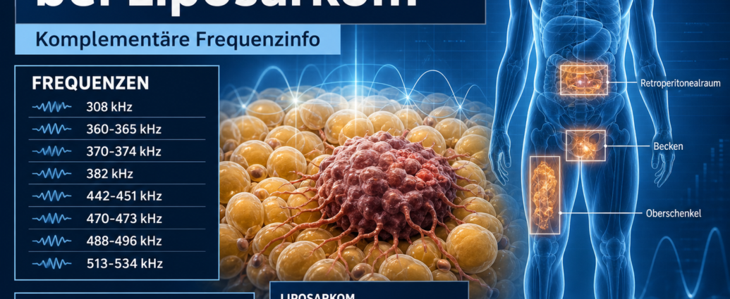
Frequency Therapy for Liposarcoma
308 kHz,
360–365 kHz,
370–374 kHz,
382 kHz,
442-451 kHz,
470–473 kHz,
488–496 kHz,
513–534 kHz.
These frequencies are described in the literature as common resonances associated with liposarcoma. In the context of frequency therapy, they can be used as a supplementary guide for documentation, resonance analysis, and individualized treatment.
Frequency Information: Viral and Mycoplasmal Resonance Patterns
In the literature, liposarcoma is associated with viral and mycoplasmal co-infections. Specific mention is made of sarcoma viruses, human lymphotropic viruses, and Mycoplasma fermentans.
Frequency Therapy for Concomitant Conditions
Sarcoma viruses,
human lymphotropic viruses,
Mycoplasma fermentans,
secondary concomitant infections.
These accompanying patterns are considered in a complementary context alongside adipose tissue, the tumor microenvironment, the immune status, and individual regulatory capacity.
Frequency Therapy for Liposarcoma: Comparison of Frequency Patterns
When comparing the frequencies, it is noticeable that lipomas and liposarcomas share common frequency ranges. In particular, the 442–451 kHz range is mentioned for both. The ranges around 308 kHz and 360–365 kHz also show similarities between benign and malignant changes in adipose tissue.
In the case of liposarcoma, additional resonance fields are present:
370–374 kHz,
382 kHz,
470–473 kHz,
488–496 kHz,
513–534 kHz.
In a complementary context, these additional ranges can be viewed as an extension of the resonance pattern observed in malignant adipose tissue tumors. In particular, the 488–496 kHz and 513–534 kHz ranges form higher-frequency bands that are not mentioned in this context in relation to lipomas.
Frequency Therapy for Liposarcoma: Summary
Liposarcoma is a malignant tumor of adipose tissue and is one of the most common soft tissue sarcomas in adults. It frequently occurs in deep soft tissue compartments, particularly on the thigh, in the buttocks, on the leg, in the shoulder region, and in the posterior abdomen.
In conventional medicine, a distinction is made between well-differentiated, myxoid, round-cell, pleomorphic, and mixed-type liposarcomas. Diagnosis is made through imaging, biopsy, histological examination, and, if necessary, molecular genetic analysis. The primary treatment is usually surgical removal.
Frequency therapy offers a complementary perspective. The literature cites the following resonance frequencies for liposarcomas: 308 kHz, 360–365 kHz, 370–374 kHz, 382 kHz, 442–451 kHz, 470–473 kHz, 488–496 kHz, and 513–534 kHz. In addition, lipoma frequencies such as 308–312 kHz, 360–364 kHz, and 442–451 kHz are listed for comparison. These frequency lists can be used in a complementary context for documentation, resonance analysis, and individualized frequency therapy work.

Comments are closed, but trackbacks and pingbacks are open.